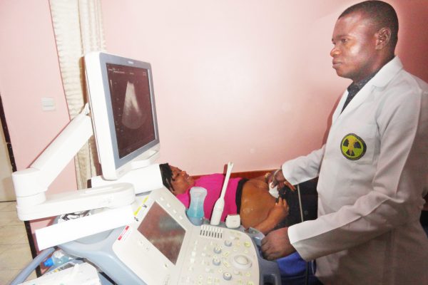
- ekene
- 0 Comments
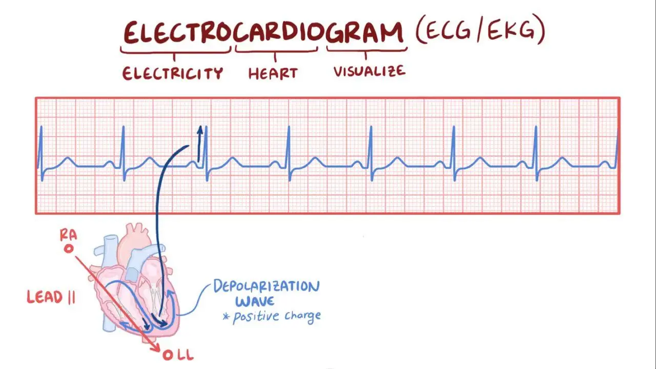
Electrocardiography is the process of producing an electrocardiogram (ECG ), a recording of the heart’s electrical activity. It is used to check your heart’s rhythm and electrical activity.
There are three main components to an ECG: the P wave, which represents depolarization of the atria; the QRS complex, which represents depolarization of the ventricles; and the T wave, which represents repolarization of the ventricles.
WHY DO AN ECG
- Chest pain or suspected myocardial infarction (heart attack),
- Evaluate shortness of breath, fainting, seizures, severe tiredness, fainting or arrhythmias including new onset palpitations
- To identify irregular heartbeats
- Monitoring of known cardiac arrhythmias
- Electrolyte abnormalities, such as hyperkalemia
- Cardiac stress testing
- To help determine the overall health of the heart before procedures such as surgery; or after treatment for conditions such as a heart attack (myocardial infarction, or MI), endocarditis (inflammation or infection of one or more of the heart valves); or after heart surgery or cardiac catheterization
- To see how an implanted pacemaker is working
- To determine how well certain heart medicines are working
- To get a baseline tracing of the heart’s function during a physical exam; this may be used as a comparison with future ECGs, to determine if there have been any changes

TYPES OF ECG
- 12 Lead/Normal ECG
- Stress ECG
- HOLTER ECG
12 LEAD ECG
A 12-lead electrocardiogram (ECG) is a medical test that is recorded using leads, or nodes, attached to the body. Electrocardiograms, sometimes referred to as ECGs, capture the electrical activity of the heart and transfer it to graphed paper. The results can then be analyzed by medical professionals, such as cardiologists, cardiac nurses and technicians.
STRESS ECG
A cardiac stress test (also referred to as a cardiac diagnostic test, cardiopulmonary exercise test, or abbreviated CPX test) is a cardio logical test that measures the heart’s ability to respond to external stress in a controlled clinical environment.
HOW ECG IS CARRIED OUT
Leads and electrodes are placed in the limbs and chest respectively. The lead is placed from the anterior right mid clavicular line to the left mid auxiliary line.
V1 is placed on the fourth intercostal space.
V2 is placed on the 4th intercostal space to the left sternum.
V3 is placed directly between the leads V2 and V1.
V4 is placed on the 5th intercostal space of the mid clavicular line.
V5 is level with V4 at the left anterior axillary line.
V6 is level with V5 at the mid-axillary line (directly under the midpoint of the armpit).
12 Lead resting ECG electrode placement
RA – Right forearm or wrist
LA – Left forearm or wrist
LL – left lower leg proximal to ankle
RL – Right lower leg proximal to ankle
Afterwards the patient’s bio data weight and height is entered on the machine. The machine then produces a chart which will be used by the cardiologist for reporting.
INTERPRETATION OF ECG
An ECG is reported by a Cardiologist
Where can I carry out ECG
At Lifebridge Medical Diagnostics Center we carry out all kinds of ECG scan at an affordable price. You can visit us at No 15a Yawuri Street behind Rita Lori hotel Garki 2, Abuja. Or call 08180626274
